
researchers of the University of La Laguna (ULL) participate in a study on the deformations found in bones of dinosaurs sauropods of Argentinawhich could be due to accidents suffered by these animals or even to their movement during copulation.
In recent decades, access to different medical technologies, such as tomography, has allowed paleontologists to carry out new research on the paleobiology of dinosaurs with which to answer numerous questions related not only to their biology, but also to their behavior.
The study of the paleopathologies of fossil organisms also allows us to know aspects of their life, such as the social interactions between individuals of the same species, of different species or even with the same environment in which they lived, the University of La Laguna said this Wednesday. (ULL) in a statement.
For this reason, the researcher of the Department of Animal Biology, Pedology and Geology of the ULL Penélope Cruzado-Caballero, together with Leonardo Filippi, from the Museo Municipal Argentino Urquiza de Neuquén; Javier González-Dionis from Conicet IPGP-CENPAT and Ignacio Canudo from the University of Zaragoza studied two tail vertebrae belonging to titanosaur sauropod dinosaurs.
These skeletal remains were found in the towns of La Invernada and Loma de los Jotes, near the city of Rincón de los Sauces (Neuquén, Argentina).
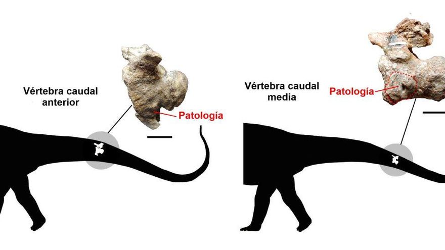
What caught our attention was that they presented certain deformations and thickenings that were not typical of the normal anatomy of these vertebrae, which made the research team think about the possible presence of some type of pathology that the dinosaurs to which these belonged would have suffered. fossils.
To study these strange structures that the vertebrae presented, a computerized tomography was performed at the “Eva Perón” Clinic and Maternity Hospital in Rincón de los Sauces (Neuquén, Argentina).
After an analysis of the surface of the fossils and the images obtained with the tomography, the paleontologists discovered that one of the vertebrae presented a pathology that was difficult to appreciate, located between two bones: the ventral part of the body of the vertebra and a bone called chevron that articulates with it.
Between these two bones, an amorphous mass of bone was observed that covered them, which was interpreted as a spondyloarthropathy that could have been produced by different causes.
In this case, this type of arthropathy (joint disease) would not have caused pain or reduced mobility of the tail, contrary to what must have occurred with the second vertebra studied.
This is a vertebra from the middle part of the tail of another dinosaur, which presented a bone growth on one of its sides that is clearly different from the rest of the vertebra.
In addition, associated with this bone growth, a hole was observed that enters the bone and has the typical shape of what is known as a cloaca. This hole is a structure that occurs when there is an infection in the bone, which produces pus and needs to be expelled from it.
This type of infection that affects the bones is known as osteomyelitis and due to its location and the development of the infection, it was concluded that it could have caused the dinosaur some discomfort and pain, and could even have restricted some degree of mobility of the posterior part of the tail.
Along with the “paleoveterinary” work carried out, an exhaustive analysis was also made of everything that has been published worldwide on the pathologies recorded in the tails of sauropod dinosaurs, with the aim of seeing if there is one that affects more than another. in this group of animals.
As a result, it was seen that most of the works on sauropods were in the group of titanosaurs (69% of the total) and that, in terms of the diversity of pathologies that affected sauropods, the one known as DISH (Skeletal Hyperostosis) stood out. diffuse idiopathic with 36% of cases) or the already mentioned spondyloarthropathy (16% of cases).
As a curiosity, the ULL indicates that it has been proposed that these pathologies could be related to the moment of copulation, since they could be the result of the reaction of the bone to stress microfractures resulting from an upright posture of the animal that would support all its weight on the ground between the hind legs and tail.


